Home
/ Sketch And Label Of A Cross Section Of A Long Bone : Draw The Given Diagram And Label The Following Parts A Spongy Boneb Periosteumc Yellow Marrowd Compact Bone - The diaphysis and the epiphysis.
Sketch And Label Of A Cross Section Of A Long Bone : Draw The Given Diagram And Label The Following Parts A Spongy Boneb Periosteumc Yellow Marrowd Compact Bone - The diaphysis and the epiphysis.
Sketch And Label Of A Cross Section Of A Long Bone : Draw The Given Diagram And Label The Following Parts A Spongy Boneb Periosteumc Yellow Marrowd Compact Bone - The diaphysis and the epiphysis.. A long bone has two parts: Then, fill in the table below to describe each. A typical long bone shows the gross anatomical characteristics of bone. Sketch and label of a cross section of a long bone / 1: Learners should accurately draw a long bone, resembling that in figure 6.24.
Wrist cross section educational structure scheme vector illustration. Sketch and label of a cross section of a long bone / 1: Create a drawing of the bone section in your laboratory journal and label the areas listed above. Cartilaginous area at the ends of long bones where lengthwise growth takes place in the immature skeleton. Proximal epiphysis, distal epiphysis, diaphysis, metaphysis, medullary cavity, epiphyseal line 2.
Diagram Of A Longitudinal Section Of A Long Bone Stock Illustration Download Image Now Istock from media.istockphoto.com Schematic drawing of a longitudinal section through a long bone. This is the long central shaft. Sketch and label of a cross section of a long bone. A long bone has a shaft and 2 ends. Create a drawing of the bone section in your laboratory journal and label the areas listed above. Osteons are oriented parallel to the diaphysis of the long bone. This photo shows a cross section through bone. In the space provided draw a longitudinal section of a long bone and label the following parte proximal epiphysis, distal epiphysis, diaphysis, metaphysis, medullary cavity, epiphyseal line 2.
The periosteum contains many strong collagen fibers that are used to firmly anchor tendons and muscles to the bone for movement.
Complete figure 6.1a by labeling compact bone and spongy bone. Once we stop growing (between 18. In the space provided, draw a longitudinal section of a long bone and label the following parts: Label lines should not cross ; Bone matrix and cells bone matrix osseous tissue is a connective tissue and like all connective tissues contains relatively few cells and large amounts of extracellular matrix. The humerus is the long bone in the upper arm. Area between the diaphysis and epiphysis at both ends of the bone. Cross section of long bone. Label the haversian canal, osteocyte (mature bone cell) in lacuna, and canaliculi. Bring your designs to life with branding, web, mobile, and print mockups in various styles. The ends of a long bone contain spongy bone and an epiphyseal line. The diaphysis and the epiphysis. Create a drawing of the bone section in your laboratory journal and label the areas listed above.
At the elbow, it connects primarily to the ulna, as the forearm's radial bone connects to the. (do not copy and paste a picture from the text or internet.) A long bone has two parts: Bone test anatomy and physiology 12 photos of the bone test anatomy and physiology anatomy and physiology bone lab test, anatomy and physiology bone markings test, anatomy and physiology bone practical test, anatomy and physiology bone tissue test, anatomy and physiology test on bone tissue, bone, anatomy and. The periosteum contains many strong collagen fibers that are used to firmly anchor tendons and muscles to the bone for movement.
Bone Human Anatomy from theodora.com Related posts of cross section of human bone diagram bone in arm pictures. (do not copy and paste a picture from the text or internet.) As the names suggest compact bone looks compact and the spongy bone looks like sponges. Make a pencil sketch and use markers or colored pencils to add details. The structure of a long bone allows for the best visualization of all of the parts of a bone ( figure 6.7 ). Sketch and label of a cross section of a long bone / 1: The central haversian canal, and horizontal canals (perforating/ volkmann's) canals contain blood vessels and nerves from the periosteum. This is the long central shaft.
The diaphysis of a long bone is composed of bone tissue while the epiphysis is made of 3.
Use colored pencils to draw and label the following structures as they appear using the 40x objective. Sketch and label of a cross section of a long bone / 1: Sketch and label of a cross section of a long bone. Schematic drawing of a longitudinal section through a long bone. Cow and human long bones have a similar general structure. (do not copy and paste a picture from the text or internet.) Create a drawing of the bone section in your laboratory journal and label the areas listed above. The humerus is the long bone in the upper arm. Growth in length of a bone occurs at the 4. External circumferential lamellae, osteon, central canal, perforating canals, lacuna, canaliculi, concentric lamellae. Chapter 6 bones and skeletal tissues flashcards quizlet. The structure of a long bone allows for the best visualization of all of the parts of a bone (figure 1). The diaphysis of a long bone is composed of bone tissue while the epiphysis is made of 3.
Create a drawing of the bone section in your laboratory journal and label the areas listed above. Schematic drawing of a longitudinal section through a long bone. Bone remodeling and repair 11. Describe how this is live tissue which is both strong and slightly flexible osteons are the structural unit of compact bone, and enable to the bone to be able to bare weight with the way they are structured. Structure of a long bone , section, human body, drawing.
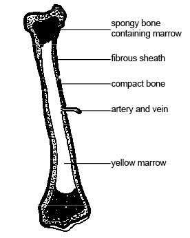
Anatomy And Physiology Of Animals The Skeleton Wikibooks Open Books For An Open World from upload.wikimedia.org Sketch and label of a cross section of a long bone. Long bones have a thick outside layer of compact bone and an inner medullary cavity containing bone marrow. The structure of a long bone allows for the best visualization of all of the parts of a bone ( figure 6.7 ). Sketch and label of a cross section of a long bone : Label the parts of a long bone. Bone test anatomy and physiology 12 photos of the bone test anatomy and physiology anatomy and physiology bone lab test, anatomy and physiology bone markings test, anatomy and physiology bone practical test, anatomy and physiology bone tissue test, anatomy and physiology test on bone tissue, bone, anatomy and. Describe how this is live tissue which is both strong and slightly flexible osteons are the structural unit of compact bone, and enable to the bone to be able to bare weight with the way they are structured. Then, fill in the table below to describe each.
End of a long bone.
The structure of a long bone allows for the best visualization of all of the parts of a bone ( figure 6.7 ). As the names suggest compact bone looks compact and the spongy bone looks like sponges. Sketch and label of a cross section of a long bone / 1: Diaphysis • shaft of the long bone. Cow and human long bones have a similar general structure. This is the long central shaft. Draw and label the following structures as they appear using the 10x objective o bone marrow o bony trabeculae activity 5.2.3: You can specify conditions of storing and accessing cookies in. Related posts of cross section of human bone diagram foot bone anatomy x ray. The end of a growing tibia, cut lengthwise*. Smartdraw includes 1000s of professional healthcare and anatomy chart templates that you can modify and make your own. The compact bone is made up of osteon. Use the internet or a reference textbook to help you identify the external features of long bone listed below.